Hemorrhagic Stroke

Introduction

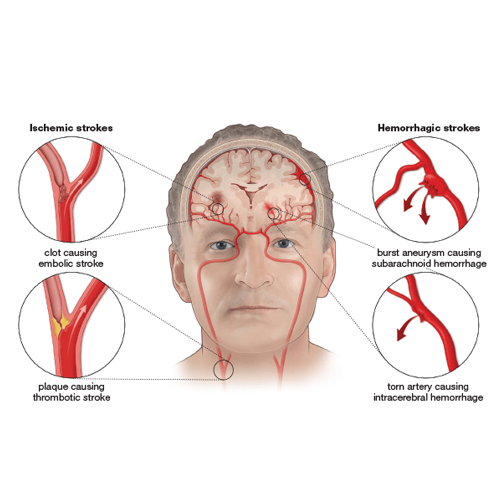
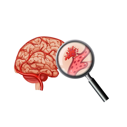
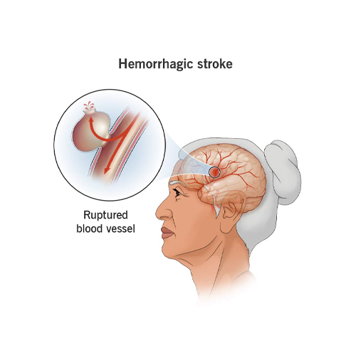
Hemorrhagic stroke is a severe medical condition that occurs when a blood vessel in the brain ruptures, leading to bleeding within or around the brain. This condition accounts for approximately 13% of all strokes and is associated with high morbidity and mortality rates.

Hemorrhagic stroke is classified into two main types: intracerebral hemorrhage (ICH) and subarachnoid hemorrhage (SAH). Understanding the causes, symptoms, diagnosis, and treatment of hemorrhagic stroke is essential for improving patient outcomes and reducing the risk of complications.

Types of Hemorrhagic Stroke

1. Intracerebral Hemorrhage (ICH)

  • ✅ This occurs when an artery in the brain bursts, causing blood to leak into the surrounding brain tissue.
  • ✅ The bleeding increases pressure in the brain and can damage brain cells.
  • ✅ Common causes include hypertension, head trauma, blood vessel abnormalities, and blood-thinning medications.

2. Subarachnoid Hemorrhage (SAH)

  • ✅ This occurs when there is bleeding into the space between the brain and the thin tissues covering it (subarachnoid space).
  • ✅ The most common cause is the rupture of an aneurysm, a weakened blood vessel that bulges and bursts.
  • ✅ Other causes include arteriovenous malformations (AVMs) and head injuries.

Causes and Risk Factors

Several factors contribute to the development of hemorrhagic stroke, including:
  • ✔️ Hypertension (High Blood Pressure): The leading cause of ICH, chronic high blood pressure weakens blood vessels, making them prone to rupture.
  • ✔️ Aneurysms: These are balloon-like bulges in blood vessels that can rupture and lead to SAH.
  • ✔️ Arteriovenous Malformations (AVMs): Abnormal tangles of blood vessels in the brain that can rupture.
  • ✔️ Head Trauma: Injuries can cause bleeding in or around the brain.
  • ✔️ Blood Thinners: Medications such as anticoagulants (e.g., warfarin, heparin) increase the risk of bleeding.
  • ✔️ Liver Disease: Impairs blood clotting, increasing the likelihood of hemorrhage.
  • ✔️ Smoking and Alcohol Use: Both can contribute to high blood pressure and weakened blood vessels.
  • ✔️ Genetic Factors: A family history of stroke, aneurysms, or AVMs can increase risk.
  • ✔️ Age and Gender: Older adults are at higher risk, and women may have a slightly increased risk of SAH compared to men.

Symptoms of Hemorrhagic Stroke

Symptoms of a hemorrhagic stroke develop suddenly and can include:
  • ✔️ Sudden, severe headache (often described as "the worst headache of my life")
  • ✔️ Nausea and vomiting
  • ✔️ Weakness or numbness on one side of the body
  • ✔️ Vision disturbances
  • ✔️ Difficulty speaking or understanding speech
  • ✔️ Loss of coordination and balance
  • ✔️ Seizures
  • ✔️ Loss of consciousness
  • ✔️ Sensitivity to light (photophobia) in cases of SAH

Diagnosis

Diagnosing hemorrhagic stroke requires rapid medical evaluation, including:

Physical and Neurological Examination

  • ✔️ Assessment of symptoms
  • ✔️ Testing reflexes, muscle strength, and coordination

Imaging Tests

  • ✔️ Computed Tomography (CT) Scan: The most common and quickest test to detect bleeding in the brain.
  • ✔️ Magnetic Resonance Imaging (MRI): Provides detailed images of brain structures and can identify smaller bleeds.
  • ✔️ Cerebral Angiography: Used to visualize blood vessels and detect aneurysms or AVMs.

Blood Tests

  • ✔️ Evaluate clotting function
  • ✔️ Check platelet levels
  • ✔️ Detect other factors that may contribute to bleeding.

Lumbar Puncture

  • ✔️ In cases where SAH is suspected but not visible on a CT scan.
  • ✔️ A spinal tap may be performed to detect blood in cerebrospinal fluid.

Treatment Options

Treatment depends on the severity, location, and cause of the hemorrhage. Major approaches include:

Emergency Treatment

    Stabilization:
  • Patients may require oxygen, IV fluids, and blood pressure management to prevent further damage.
  • Medications:
  • ✔️ Drugs to control blood pressure (e.g., labetalol, nicardipine).
  • ✔️ Medications to reduce brain swelling (e.g., mannitol, hypertonic saline).
  • ✔️ Reversal agents for blood thinners (e.g., vitamin K, prothrombin complex concentrate for warfarin reversal).
  • ✔️ Anticonvulsants to prevent seizures.

Surgical Treatment

  • ✔️ Craniotomy & Hematoma Removal: In cases of large hematomas, surgery may be required to remove the clot and relieve pressure on the brain. A craniotomy involves temporarily removing a portion of the skull to access the hematoma, stop the bleeding, and remove damaged tissue. This procedure is essential for preventing further neurological deterioration and reducing intracranial pressure.
  • ✔️ Aneurysm Clipping or Coiling: If an aneurysm is the cause, neurosurgeons may perform microsurgical clipping, where a small metal clip is placed at the base of the aneurysm to prevent further bleeding. Alternatively, endovascular coiling involves inserting a catheter through an artery and deploying small platinum coils into the aneurysm, promoting clotting and sealing off the aneurysm.
  • ✔️ AVM Removal: If an arteriovenous malformation is present, different surgical techniques may be used depending on its size and location. Options include microsurgical resection to remove the AVM completely, stereotactic radiosurgery (such as Gamma Knife) to deliver targeted radiation, or endovascular embolization, where a catheter is used to inject a substance that blocks abnormal blood flow, reducing the risk of rupture.
  • ✔️ Ventriculostomy: A procedure to drain excess cerebrospinal fluid (CSF) and reduce intracranial pressure. A catheter is placed into the brain’s ventricles to remove accumulated fluid, which can relieve pressure and prevent further damage. In cases of severe hydrocephalus, a long-term shunting system (such as a ventriculoperitoneal shunt) may be necessary to continuously divert CSF.

Rehabilitation and Recovery

Rehabilitation is crucial for stroke survivors and focuses on regaining lost abilities and adapting to new challenges.

Staging of Rehabilitation

Heidelberg Model of Rehabilitation:

    ✔️ Phase I: Inpatient Rehabilitation
  • Early mobilization and self-care training.
  • Prevention of complications during the acute phase.
  • Intensive physiotherapy and occupational therapy to restore motor functions.
  • ✔️ Phase II: Early Outpatient Rehabilitation
  • Structured therapy programs, including exercise and patient education.
  • Supervised physical training to enhance mobility and coordination.
  • Psychological support and cognitive rehabilitation for mental recovery.
  • ✔️ Phase III: Long-Term Rehabilitation
  • Community-based programs for continuous support and secondary prevention.
  • Adaptive strategies for daily living and workplace reintegration.
  • Tele-rehabilitation services for remote monitoring and therapy.

Bavarian Rehabilitation Techniques

1. Klinik Bavaria Kreischa

  • ✔️ Advanced physiotherapy focusing on muscle re-education and neuroplasticity.
  • ✔️ Aquatic therapy for improved balance, strength, and endurance.
  • ✔️ Specialized speech and language therapy for post-stroke communication difficulties.

2. Klinik Bavaria Bad Kissingen

  • ✔️ Neurological rehabilitation incorporating movement training and robotic-assisted therapy.
  • ✔️ Cognitive therapy to enhance memory, attention, and problem-solving skills.
  • ✔️ Comprehensive interdisciplinary approaches integrating neurology, physiotherapy, and occupational therapy.

Innovative Rehabilitation Technologies:

Robotic-Assisted Therapy:

  • ✔️ Exoskeletons and robotic arms assist in movement recovery by providing precise, controlled motions for patients with motor impairments.
  • ✔️ Robotic gait trainers help stroke survivors regain walking ability through repetitive training and muscle stimulation.

Electromagnetic Stimulation:

  • ✔️ Transcranial Magnetic Stimulation (TMS) is used to stimulate neural pathways and promote brain plasticity.
  • ✔️ Functional Electrical Stimulation (FES) applies controlled electrical impulses to weak muscles, aiding in movement restoration.

Virtual Reality Therapy:

  • ✔️ Engages patients in interactive, immersive environments to enhance motor and cognitive rehabilitation.
  • ✔️ Improves coordination, balance, and neuroplasticity by simulating real-life tasks.

Brain-Computer Interfaces (BCIs):

  • ✔️ Allows patients to control assistive devices using brain activity, providing communication and mobility solutions for those with severe impairments.
  • ✔️ Helps retrain brain functions by translating neural signals into physical actions, aiding in movement rehabilitation.

Conclusion

Hemorrhagic stroke is a life-threatening condition that requires immediate medical attention. Advances in treatment and rehabilitation have improved outcomes, but prevention remains crucial. Early intervention, comprehensive rehabilitation, and lifestyle modifications are key to recovery and reducing the risk of future strokes.