Subscribe to out newsletter today to receive latest news administrate cost effective for tactical data.
2478 Street City Ohio 90255
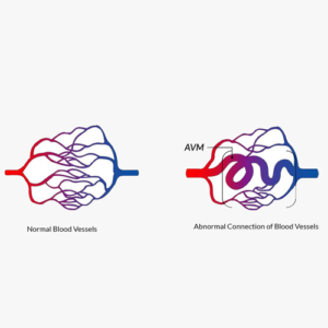
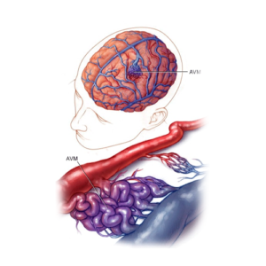
Aneurysms and arteriovenous malformations (AVMs) are serious neurovascular conditions that can lead to life-threatening complications such as hemorrhagic stroke and brain damage.
Germany is at the forefront of neurovascular care, offering advanced diagnostic tools, minimally invasive surgical techniques, and world-class rehabilitation programs. At WEGOVITA, we connect patients with Germany’s leading neurosurgeons, interventional radiologists, and rehabilitation experts to ensure the best possible treatment outcomes.
A. Primary Causes
Aneurysm Formation:
Hypertension (High Blood Pressure) – Increased pressure weakens artery walls.
Atherosclerosis – Plaque buildup damages arteries, increasing risk.
Genetic Disorders – Conditions like Ehlers-Danlos and Marfan syndrome can cause vascular weakness.
Head Trauma – Can lead to dissecting aneurysms.
Infections (Mycotic Aneurysm) – Bacterial or fungal infections weakening artery walls.
AVM Development:
Congenital Factors – AVMs typically form during fetal development.
Genetic Mutations – Associated with hereditary hemorrhagic telangiectasia (HHT).
Unknown Causes – Some AVMs arise sporadically without a clear genetic link.
B. Risk Factors
Aneurysm Formation:
Modifiable Risk Factors:
Hpyertension – Major risk factor for aneurysm rupture.
Smoking – Weakens arterial walls and increases rupture risk.
Heavy Alcohol Consumption – Can lead to blood vessel abnormalities.
llicit Drug Use (Cocaine, Amphetamines) – Causes vascular stress and rupture risk.
Obesity & Sedentary Lifestyle – Increases cardiovascular risks.
Non-Modifiable Risk Factors:
✅ Family History – Increased risk if a close relative has an aneurysm or AVM.
✅ Age & Gender – Aneurysms are more common in people over 40 and women.
Unruptured Aneurysm Symptoms:
Persistent headaches
Blurred or double vision
Pupil dilation
Numbness or weakness in one side of the face
Speech difficulties
Ruptured Aneurysm Symptoms (Medical Emergency):
Sudden, severe headache (“thunderclap headache”)
Nausea and vomiting
Stiff neck and sensitivity to light
Loss of consciousness or coma
Seizures
AVM Symptoms:
Seizures
Headaches
Muscle weakness or numbness
Vision problems
Cognitive or speech difficulties
Germany provides cutting-edge imaging technologies for aneurysm and AVM detection, ensuring precise diagnosis and treatment planning. Early and accurate detection is crucial for preventing life-threatening complications such as brain hemorrhages and seizures. The following state-of-the-art diagnostic modalities are widely used in German neurovascular centers:
Computed Tomography Angiography (CTA)– MA high-resolution imaging technique that combines traditional CT scanning with a contrast dye injection to produce detailed 3D images of blood vessels in the brain. CTA is crucial for detecting aneurysms, vascular malformations, and stenotic lesions. It provides a rapid and non-invasive method for evaluating vessel integrity and the presence of abnormal dilations or tangles.
Magnetic Resonance Angiography (MRA) – A non-invasive imaging technique using powerful magnetic fields and radio waves to create highly detailed images of cerebrovascular structures. MRA is particularly useful for detecting unruptured aneurysms and AVMs, assessing blood flow, and identifying vessel abnormalities without exposing patients to ionizing radiation. Contrast-enhanced MRA may be used for even more precise visualization.
Cerebral Angiography (Digital Subtraction Angiography – DSA) – Considered the gold standard for detailed vascular assessment, DSA provides real-time, high-resolution images of blood vessels by injecting contrast dye directly into the cerebral arteries through a catheter. This technique allows for precise visualization of aneurysms, AVMs, arterial stenosis, and other vascular anomalies. It is often used before interventional procedures such as endovascular coiling or embolization.
Transcranial Doppler Ultrasound (TCD) – A non-invasive ultrasound technique that evaluates blood flow abnormalities in the intracranial arteries. TCD helps detect vasospasm (narrowing of arteries due to aneurysm rupture), emboli, and changes in cerebral blood circulation. It is commonly used in monitoring patients post-aneurysm treatment and for assessing stroke risk in AVM patients.
Electroencephalography (EEG) – A diagnostic test that measures electrical activity in the brain to detect seizures, which are common in patients with AVMs. EEG can help identify abnormal brain wave patterns, guide seizure management strategies, and assess the impact of AVMs on neurological function. Continuous or prolonged EEG monitoring may be recommended for patients with recurrent seizures.
Germany’s advanced neurovascular diagnostics ensure early detection, precise treatment planning, and optimal patient outcomes, leveraging the latest in medical imaging technology to provide world-class care.
A. Aneurysm Treatment
Endovascular Coiling (Minimally Invasive):
A catheter places platinum coils inside the aneurysm to prevent rupture.
Effective for small to medium-sized aneurysms.
Surgical Clipping:
Open brain surgery to clip off the aneurysm from blood circulation.
Preferred for large or complex aneurysms.
Flow Diverter Stents:
Diverts blood flow away from the aneurysm, promoting clot formation.
Anti-Hypertensive & Lifestyle Management:
Medications to control blood pressure and prevent rupture.
B. AVM Treatment
Stereotactic Radiosurgery (Gamma Knife, CyberKnife):
High-dose radiation targets AVM to shrink it over time.
Ideal for small or deep-seated AVMs.
Endovascular Embolization:
Catheter-based procedure injecting glue-like material to block abnormal vessels.
Often a pre-surgical or adjunctive treatment.
Microsurgical Resection:
Complete surgical removal of the AVM.
Best for accessible and high-risk AVMs.
Seizure & Symptom Management:
Anti-epileptic drugs for seizure control.
Stage 1: Acute Neurorehabilitation (0-2 Weeks)
The acute phase of neurorehabilitation focuses on stabilizing the patient’s condition, preventing complications, and initiating early recovery strategies. During the first two weeks, intensive care teams work to optimize neurological function, minimize disability, and create a personalized rehabilitation plan tailored to the patient’s needs.
Early Mobilization – Early movement is encouraged within the first 24-48 hours after surgery or hemorrhagic event to prevent complications such as deep vein thrombosis (DVT), pneumonia, and muscle atrophy. Physical therapists assist with passive and active range-of-motion exercises, sitting balance training, and gradual standing and walking to promote circulation and functional recovery.
Swallowing & Speech Therapy – Many patients experience swallowing difficulties (dysphagia) or speech impairments due to brain injury or post-surgical effects. A specialized speech-language pathologist (SLP) assesses swallowing function to prevent aspiration pneumonia and choking risks. Speech therapy focuses on articulation, voice control, and cognitive-linguistic skills to improve communication.
Cognitive Assessments & Neurological Evaluations – A multidisciplinary team, including neurologists and neuropsychologists, conducts comprehensive cognitive assessments to identify deficits in memory, attention, problem-solving, and executive functions. These evaluations help guide therapy plans and predict recovery outcomes.
Pain Management & Spasticity Control – Post-surgical or post-stroke pain, muscle stiffness, and spasticity are common challenges during acute rehabilitation. Treatment includes medications, nerve blocks, physical therapy, and in some cases, botulinum toxin injections to manage muscle tightness.
Nutritional Support & Hydration Therapy – Proper nutrition is essential for brain recovery. Dietitians create customized dietary plans to support healing and prevent malnutrition, which can delay recovery. Patients who have difficulty swallowing may receive modified diets or temporary feeding tube support.
Family & Caregiver Education – During this phase, healthcare providers educate family members about the patient’s condition, expected progress, and how to assist in basic care needs. Psychological support is also offered to caregivers to help them cope with the emotional impact of their loved one’s condition.
During this critical phase of recovery, patients continue their rehabilitation efforts with a more intensive, structured approach to restore lost functions, improve mobility, and enhance independence. This stage focuses on physical recovery as well as cognitive and emotional well-being.
Physical & Occupational Therapy – This phase emphasizes strengthening motor function, improving coordination, and increasing endurance. Physical therapy includes gait training, balance exercises, and strength conditioning, while occupational therapy focuses on daily living skills, fine motor tasks, and adaptive techniques to regain independence in personal care and work-related activities.
Speech & Language Therapy – Many patients experience speech difficulties, cognitive impairments, or swallowing issues after an aneurysm or AVM-related event. Therapy sessions focus on articulation, comprehension, and speech production exercises. Swallowing assessments and exercises help prevent aspiration pneumonia and ensure safe eating and drinking.
Psychological Counseling & Emotional Support – Coping with the aftermath of an aneurysm or AVM can be emotionally challenging. Patients may experience anxiety, depression, or frustration due to their condition. Psychological counseling, cognitive behavioral therapy (CBT), and support groups help patients manage stress, build resilience, and maintain a positive outlook during recovery.
Cognitive Rehabilitation Therapy (CRT) – Patients who have suffered from brain hemorrhages or neurological deficits may need cognitive training to enhance memory, problem-solving abilities, and attention span. CRT includes exercises, computer-based training, and structured cognitive tasks aimed at improving executive functioning and mental agility.
Pain Management & Spasticity Control – Some patients may experience muscle stiffness, spasticity, or neuropathic pain during recovery. Rehabilitation specialists use medications, botulinum toxin injections, massage therapy, and stretching exercises to alleviate discomfort and improve mobility.
Nutritional Counseling & Dietary Support – Proper nutrition plays a crucial role in the recovery process. Dietitians work with patients to optimize their diet, ensuring adequate intake of essential nutrients that promote brain healing, reduce inflammation, and support overall health. Special attention is given to dietary modifications for those with swallowing difficulties.
Family & Caregiver Education – Equipping caregivers with the right knowledge and skills is essential for a patient’s long-term recovery. Caregivers receive training on assisting with mobility, communication strategies, recognizing warning signs of complications, and providing emotional support.
This phase of rehabilitation sets the foundation for long-term recovery, helping patients regain confidence and independence in their daily lives.
Germany provides cutting-edge imaging technologies for aneurysm and AVM detection, ensuring precise diagnosis and treatment planning. Early and accurate detection is crucial for preventing life-threatening complications such as brain hemorrhages and seizures. The following state-of-the-art diagnostic modalities are widely used in German neurovascular centers:
Computed Tomography Angiography (CTA) – A high-resolution imaging technique that combines traditional CT scanning with a contrast dye injection to produce detailed 3D images of blood vessels in the brain. CTA is crucial for detecting aneurysms, vascular malformations, and stenotic lesions. It provides a rapid and non-invasive method for evaluating vessel integrity and the presence of abnormal dilations or tangles.
Magnetic Resonance Angiography (MRA) – A non-invasive imaging technique using powerful magnetic fields and radio waves to create highly detailed images of cerebrovascular structures. MRA is particularly useful for detecting unruptured aneurysms and AVMs, assessing blood flow, and identifying vessel abnormalities without exposing patients to ionizing radiation. Contrast-enhanced MRA may be used for even more precise visualization.
Cerebral Angiography (Digital Subtraction Angiography – DSA) – Considered the gold standard for detailed vascular assessment, DSA provides real-time, high-resolution images of blood vessels by injecting contrast dye directly into the cerebral arteries through a catheter. This technique allows for precise visualization of aneurysms, AVMs, arterial stenosis, and other vascular anomalies. It is often used before interventional procedures such as endovascular coiling or embolization.
Transcranial Doppler Ultrasound (TCD) – A non-invasive ultrasound technique that evaluates blood flow abnormalities in the intracranial arteries. TCD helps detect vasospasm (narrowing of arteries due to aneurysm rupture), emboli, and changes in cerebral blood circulation. It is commonly used in monitoring patients post-aneurysm treatment and for assessing stroke risk in AVM patients.
Electroencephalography (EEG) – A diagnostic test that measures electrical activity in the brain to detect seizures, which are common in patients with AVMs. EEG can help identify abnormal brain wave patterns, guide seizure management strategies, and assess the impact of AVMs on neurological function. Continuous or prolonged EEG monitoring may be recommended for patients with recurrent seizures.
Germany’s advanced neurovascular diagnostics ensure early detection, precise treatment planning, and optimal patient outcomes, leveraging the latest in medical imaging technology to provide world-class care
During the acute phase of neurorehabilitation, the primary goal is to stabilize the patient, prevent complications, and initiate early interventions that promote recovery. German neurorehabilitation centers employ a multidisciplinary approach to maximize patient outcomes.
Early Mobilization – Prevents complications such as deep vein thrombosis (DVT), pressure ulcers, and pneumonia. Mobilization may begin as early as 24-48 hours after treatment, depending on the patient’s condition. Physiotherapists assist with passive and active movement exercises to maintain circulation and prevent muscle atrophy.
Swallowing & Speech Therapy – Many patients experience difficulty swallowing (dysphagia) after aneurysm or AVM treatment. Speech therapists assess swallowing function to prevent aspiration pneumonia and provide exercises to restore speech and communication abilities.
Cognitive Assessments – Neuropsychologists evaluate memory, attention, and executive function deficits that may arise after a brain aneurysm rupture or AVM-related hemorrhage. These assessments guide the development of personalized cognitive rehabilitation programs.
Pain Management & Symptom Control – Patients may experience headaches, neuropathic pain, or muscle stiffness. Physicians provide targeted pain relief strategies, including medication, nerve blocks, and physical therapy techniques.
Nutritional Support – Proper nutrition plays a crucial role in recovery. Dietitians develop personalized meal plans to ensure adequate caloric and nutrient intake, especially for patients with swallowing difficulties.
Psychological & Emotional Support – Anxiety, depression, and post-traumatic stress disorder (PTSD) are common after a neurovascular event. Psychologists and counselors provide emotional support, coping strategies, and, if needed, medication to manage mental health symptoms.
Caregiver Education & Support – Families receive guidance on how to assist with mobility, communication, and daily activities. Training programs help caregivers understand the rehabilitation process and provide support at home.
Stage 3: Long-Term Rehabilitation (3-6 Months+)
Long-term rehabilitation focuses on enhancing independence, improving quality of life, and leveraging cutting-edge therapies to promote neuroplasticity and functional recovery. German neurorehabilitation centers integrate advanced technologies and personalized therapy programs to support long-term recovery.
Brain-Computer Interface (BCI) Therapy – Cutting-edge AI-driven technology that enables patients to control external devices, such as robotic limbs, through brain signals. BCI therapy aids in neuroplasticity and enhances motor function recovery.
Virtual Reality (VR) Therapy – Uses immersive environments to stimulate brain activity and improve coordination, balance, and cognitive functions. VR-based exercises help retrain motor pathways and enhance rehabilitation engagement.
Constraint-Induced Movement Therapy (CIMT)– A targeted approach that forces the use of the affected limb by restricting movement of the unaffected side. This therapy strengthens neural connections and improves functional use of the impaired limb.
Advanced Robotic-Assisted Therapy – Exoskeletons and robotic devices assist with walking and upper limb movements, providing repetitive and intensive rehabilitation to maximize motor recovery.
Cognitive & Memory Rehabilitation – Computer-based cognitive training programs enhance memory, problem-solving skills, and attention span, helping patients regain independence in daily activities.
Hydrotherapy & Aquatic Rehabilitation – Water-based therapy provides a low-impact environment for improving strength, coordination, and balance, reducing stress on joints and muscles.
Community Reintegration & Vocational Therapy – Helps patients transition back to work, social activities, and daily routines. Occupational therapists guide patients in adapting to their environment and regaining independence.
By integrating state-of-the-art technologies and personalized rehabilitation programs, Germany offers world-class long-term neurorehabilitation solutions, ensuring the best possible recovery outcomes for patients with aneurysms and AVMs.
WEGOVITA offers medical coordination services by connecting international patients with top hospitals and specialists across Germany. We support access to expert evaluations, facilitate treatment logistics, and present a range of available medical options.
However, WEGOVITA does not provide direct medical treatment, make medical diagnoses, or recommend specific therapies. All final medical decisions—including diagnosis, treatment planning, and cost—are made solely by licensed medical professionals after a full clinical assessment of the individual patient.
This information is provided for informational purposes, based on internationally recognized guidelines and practices used in Germany’s leading medical institutions. It is not a substitute for professional medical advice.
💡 Interested in clinical trial references, treatment innovations, or cost comparisons? Contact our medical coordination team at info@wegovita.com for personalized assistance.
Your Health. Your Journey. With WEGOVITA.